Magnetic Resonance Imaging After Brain Injury Crucial for Diagnosis
The most important read of a brain injury is the CT scan, not the MRI. The CT scan gives vital information as to whether or not surgery is necessary. But the MRI shows much more detail in the brain. When the goal is to understand the damage, the MRI is the prominent method.
Magnetic Resonance Imaging / MRI
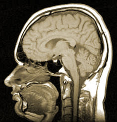
The MRI employs magnetic fields to image the brain’s cells unlike the X-ray used in the CT scan. The MRI is essentially a magnetic tunnel. It can be problematic if the patient has claustrophobia.
Benefits of the MRI rather than the CT scan:
- MRI has better resolution, better than the CT scan.
- MRI is ideal for Parenchymal Lesions. (Parenchymal Lesions are lesions to the neurons and glial cells of the brain.)
- MRI has more importance in post acute evaluations.
- MRI doesn’t use ionizing radiation, but the CT and X-ray do.
If the brain injury does not require surgery, the MRI is the best way to go. However, patients with ongoing brain damage complaints should be given an MRI on machines with the best resolution, such as the 3.0 Tesla MRI.